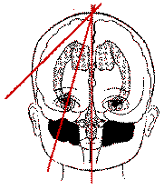
Standard cranial ultrasound scan views

Please note: images that have a white symbol at the top right, such as the Coronal views image below, indicates an image gallery that has multiple images - click on the image to open the gallery.
Introduction
A standard set of views is taken to assist with consistent visualisation of structures and in the interpretation of possible abnormalities.
The ultrasound takes a "slice" through the structure, resulting in a 2D image of a 3D structure. It is therefore important to understand the relationship of the anatomy to the image provided.
Images below detail the standard set of images taken. The accompanying images are from a 30 week infant with normal appearing anatomy.
Images are usually taken through the anterior fontanelle. The posterior fontanelle can be used if needed, and axial images are occasionally taken through the temporal bone.

In the coronal plane, a series of images are taken through the frontal lobes, more posteriorly through the ventricles and thalami, then along the plane of the choroid plexus, then superior to that.
The sagittal images are initially taken in the midline, with images then taken on both sides at the level of the lateral ventricles then periventricular areas. For the purposes of conserving space on this page, the left sagittal images have been omitted.
(Diagrams modified from Rennie JM. Neonatal cerebral ultrasound. Cambridge University Press (1997). Used with permission from the author. )
Additional images are taken according to clinical need.
Coronal views
Frontal lobes
The transducer obtains an image through the frontal lobes. The orbital ridge forms the inferior boundary of this image.
Anterior Horns of the Lateral Ventricles
The transducer is angled back. The CSF in the lateral ventricles appears as a dark image. The lateral ventricles are larger in preterm infants than in term infants. Asymmetry between the lateral ventricles is common and is not necessarily abnormal. The cavum septum pallucidum sits between the lateral ventricles and is often large in preterm infants. The corpus callosum appears above the cavum.
The Third Ventricle
With the transducer shifted slightly further back, the third ventricle appears below both lateral ventricles and the septum pallucidum. It is often small and difficult to see, but can vary considerably in size. The foramen of Monro (connecting lateral and 3rd ventricles) may be clearly seen. The brainstem may be seen as a tree-like shape.
Trigone
Angling further back cuts through the trigones of the lateral ventricles. The choroid plexus fills the lateral ventricles in this view and is prominent in preterm infants. Choroid plexus haemorrhage may be difficult to differentiate from bulky choroid. The white matter around the lateral ventricles may appear quite echodense (bright) in this plane and is sometimes called a "blush" or "flare".
Angling the transducer even more results in an image that slices above the lateral ventricles. In this plane, the occipital cortex may be visualised.
Sagittal Views
Midline Sagittal
This identifies useful landmarks. The cerebellar vermis shows up as an echogenic image in the posterior fossa. The 4th ventricle sits in front of this. The cisterna magna sits below the cerebellar vermis and is not very echogenic. The corpus callosum is seen sweeping from anterior to posterior with the cingulate gyrus above and parallel to it. The parieto-occipital sulcus is seen well above the posterior fossa.
Angled Parasagittal
The shape of the lateral ventricle is the key landmark for this view. The caudate nucleus lies below the floor of the frontal horn of the lateral ventricle; the thalamus lies behind and below it. The occipital horn of the lateral ventricle is filled with choroid plexus. The choroid tucks up in the caudothalamic groove in the floor of the lateral ventricle and may be echogenic.
Tangential Parasagittal
Further angulation of the transducer laterally results in a section lateral to the lateral ventricles. The Sylvian fissure is the key landmark in this view
